Introduction
A person with diabetes may walk into a clinic for an eye check, a kidney test, a foot assessment, or a cardiovascular review. These complications are familiar. They are written into diabetes care pathways and discussed early in the disease. But another organ is quietly involved, often without symptoms until the first fracture occurs: The Skeleton.Growing clinical evidence now shows that both type 1 and type 2 diabetes are associated with increased fracture risk, making bone health an important but often under-recognised component of diabetes care1
For many years, bone was seen mainly as a structural tissue — a mineral scaffold that holds the body upright. Diabetes, meanwhile, was understood as a disorder of glucose, insulin, blood vessels, nerves, kidneys, and the heart. The two seemed distant. Yet modern research has changed this view. Bone is now recognised as a metabolically active endocrine organ that communicates with the pancreas and other metabolic tissues through bone-derived hormones such as osteocalcin2.
This evolving understanding has brought attention to the diabetic bone paradox. In type 2 diabetes, bone mineral density may be normal or even increased, yet fracture risk remains elevated. This means that the bone may appear strong on a DXA scan but behave as fragile tissue in real life. Large cohort data published in JAMA3showed that, for a given bone mineral density or FRAX score, older adults withtype 2 diabetes had a higher risk of hip and non-spine fractures than individuals without diabetes.
The explanation lies in the difference between bone quantity and bone quality. Bone mineral density reflects how much mineral is present, but it does not fully capture collagen integrity, microarchitecture, cortical porosity, bone turnover, microdamage repair, muscle function, or fall risk. Contemporary reviews in The Lancet Diabetes & Endocrinology describe diabetic bone fragility as a complex process involving cellular dysfunction, altered bone matrix, vascular and immune changes, and musculoskeletal maladaptation to chronic hyperglycaemia4
Thus, diabetes should not be understood only as a disease of blood glucose. It is also a disease of tissue repair, endocrine signalling, vascular health, neuromuscular function, and skeletal resilience. The fracture may be the first visible clinical event, but the biological story often begins years earlier
The paradox: dense bone, fragile skeleton
A bone-density scan, usually measured by DXA, tells us how much mineral is present in bone. This is important, but it is not the whole story. Bone strength depends on both bone quantity and bone quality. Quantity refers to mineral density. Quality refers to collagen integrity, microarchitecture, turnover, repair capacity, cortical porosity, trabecular connectivity, and the ability of bone to absorb mechanical stress4,5
In type 1 diabetes, the picture is more straightforward: insulin deficiency often reduces bone formation and peak bone mass, so BMD can be low and fracture risk increases. In type 2 diabetes, the story is more paradoxical. Higher body weight and early hyperinsulinaemia may preserve or increase BMD, but chronic hyperglycaemia damages the internal material properties of bone4,5. A Lancet Diabetes & Endocrinology review describes type 1 diabetes as being associated with reduced bone mass and strength, while type 2 diabetes is characterised by increased fracture risk despite normal bone mass4
The central message is simple but clinically important:3,4
BMD is not the same as bone strength.
What glucose does to bone collagen
Bone is made of mineral crystals embedded in a collagen matrix. The mineral gives hardness; collagen gives flexibility. A healthy bone is not like chalk. It is more like reinforced biological composite material — hard enough to support weight, flexible enough to absorb impact6–8.
In chronic hyperglycaemia, glucose reacts non-enzymatically with collagen and other proteins, forming advanced glycation end products, or AGEs (Figure 1). These AGEs create abnormal cross-links within collagen. The collagen becomes stiffer, less elastic, and more brittle. The bone may still contain mineral, but its shock-absorbing capacity declines5,8.
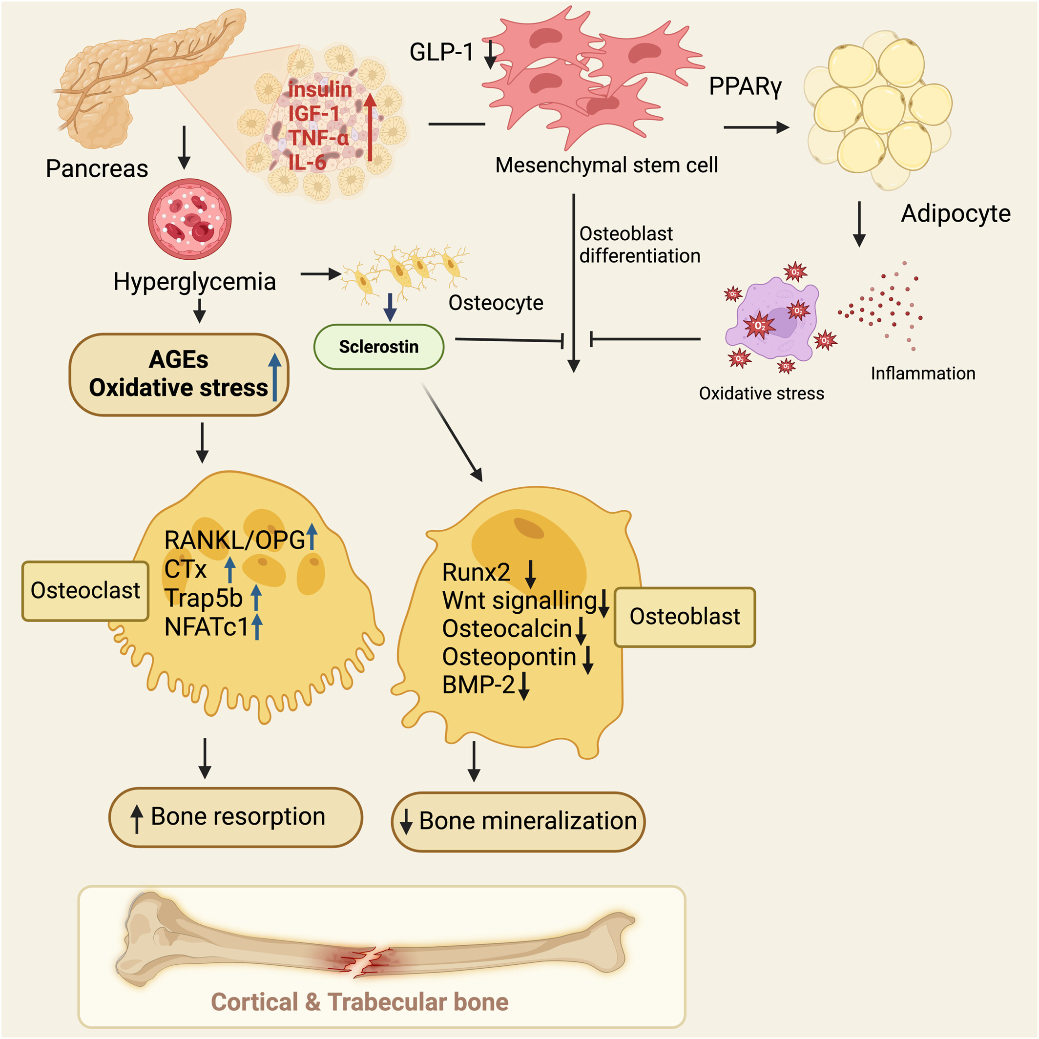
This is one of the key mechanisms of diabetic bone fragility. Reviews of fracture risk in diabetes identify AGE accumulation, low bone turnover, microstructural changes, neuropathy, hypoglycaemia, muscle weakness, visual impairment, and drug effects as contributors to fracture risk4,9.
A useful analogy is this: imagine two wooden bridges. One is slightly thinner but flexible and well maintained. The other is thick but old, dry, cracked, and poorly repaired. A scan may suggest the second bridge has more material, but it may fail sooner under stress. That is what can happen in diabetic bone.
Bone remodelling: the repair system that diabetes slows down
Bone is not static. Every day, small areas of old or damaged bone are removed and replaced. This process is called bone remodelling10.
Three main cells coordinate this system:
Osteoclasts remove old bone. Osteoblasts build new bone. Osteocytes act as sensors inside bone, detecting mechanical stress and coordinating repair11,12
In healthy physiology, this remodelling cycle clears microdamage and renews skeletal tissue. In diabetes, bone turnover is often suppressed. Osteoblast function can be impaired by hyperglycaemia, oxidative stress, inflammation, microvascular disease, and altered insulin signalling. When bone turnover slows, old glycated bone remains longer, microcracks accumulate, and skeletal resilience declines4,12,13.
This helps explain why the diabetic skeleton can be “older” than it appears on DXA. The scan measures mineral, but it does not fully measure repair quality4.
Insulin: not only a glucose hormone, but also a bone hormone
Insulin is usually introduced as the hormone that moves glucose from blood into cells. That is true, but incomplete. Insulin also acts on bone.Bone is now recognised as a metabolically active endocrine tissue that communicates with organs involved in energy balance, including the pancreas and adipose tissue14–16.
Osteoblasts have insulin receptors. When insulin binds to these receptors, it supports osteoblast activity, bone formation, and a metabolic feedback loop involving a bone-derived hormone called osteocalcin. A landmark Cell study showed that insulin signalling in osteoblasts integrates bone remodelling with whole-body energy metabolism by increasing osteocalcin activity, particularly its undercarboxylated, metabolically active form2.
This changed the way scientists viewed bone. Bone was no longer just a target of metabolic disease; it was also an active participant in metabolic regulation (Figure 2).Osteoblast-derived endocrine factors help connect bone formation with insulin production, feeding behaviour, adipose tissue metabolism, and whole-body energy balance14,16.
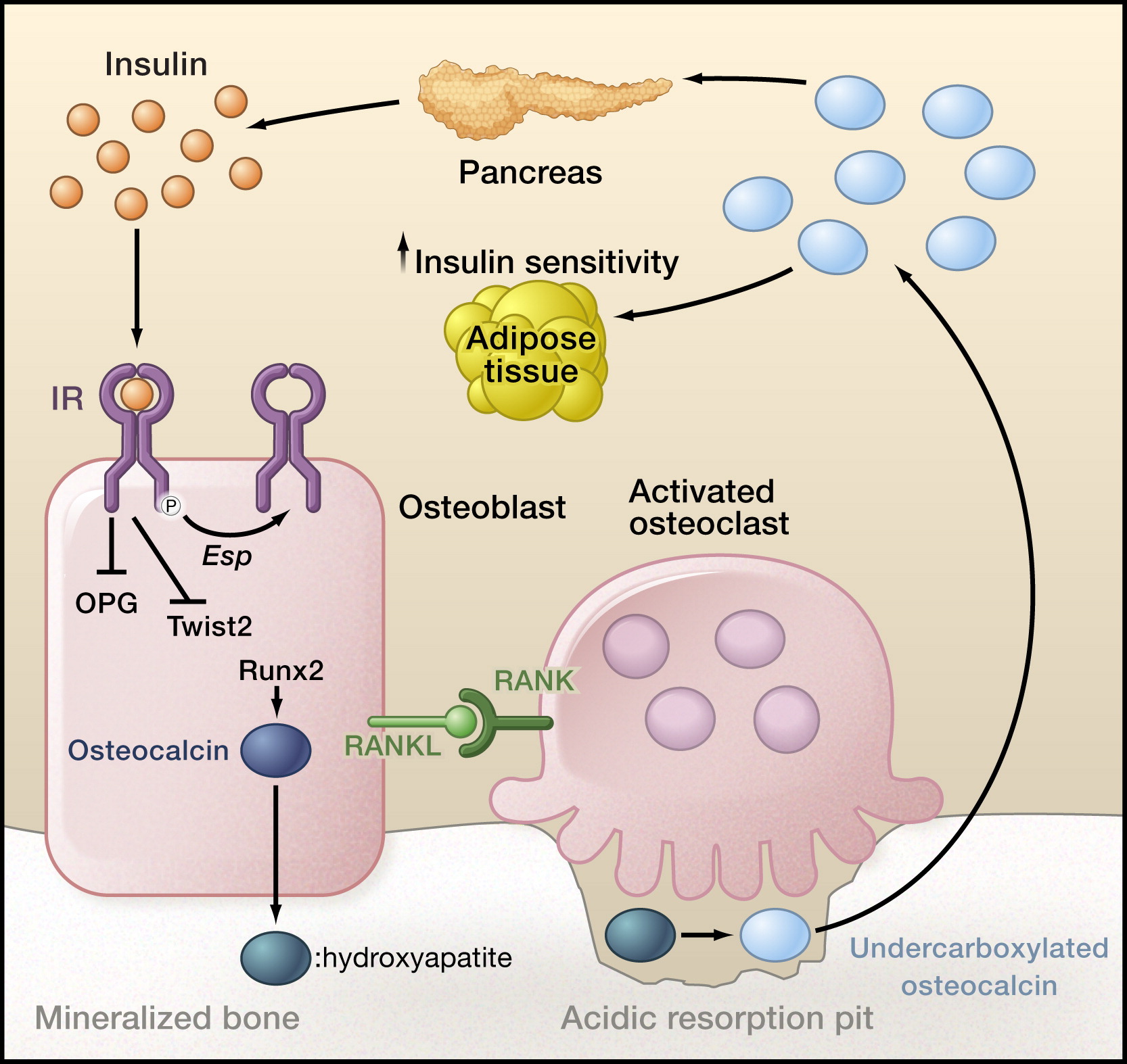
This is sometimes called the bone–pancreas endocrine loop because the pancreas signals to bone through insulin, and bone signals back to the pancreas through osteocalcin2,17.
Osteocalcin: the bone signal that talks back to metabolism
Osteocalcin is a protein made mainly by osteoblasts. Clinically, it is often considered a marker of bone formation. Biologically, it is much more interesting: it is also increasingly understood as a bone-derived endocrine signal involved in glucose and energy metabolism18–20.
Osteocalcin exists in different forms. The carboxylated form binds strongly to bone mineral and remains mainly in the bone matrix. The undercarboxylated form is more likely to enter the circulation and has stronger endocrine activity in experimental models19,21. During bone resorption, the acidic environment created by osteoclasts can promote osteocalcin decarboxylation and release, linking bone remodelling to systemic glucose metabolism2.
In animal and mechanistic studies, undercarboxylated osteocalcin can support pancreatic β-cell function, insulin secretion, insulin sensitivity, and adiponectin-related metabolic pathways2,19. Human studies are more complex, partly because assays differ and because total osteocalcin and undercarboxylated osteocalcin are not always separated. Still, the concept remains important: bone is metabolically active, and osteocalcin is one of its endocrine messengers20,21.
In diabetes, especially when bone turnover is low, osteocalcin signalling may be reduced or altered. A systematic review and meta-analysis found that people with diabetes had lower levels of bone turnover markers, including osteocalcin, compared with controls, supporting the idea that diabetes is often a state of low bone turnover22.This may weaken the normal communication between bone and glucose metabolism. The result is not merely “weak bones,” but disruption of a biological feedback system linking bone, pancreas, adipose tissue, muscle, and energy balance2,19,22.
Lipocalin-2: another bone-derived messenger
Osteocalcin is not the only metabolic signal from bone. Lipocalin-2, also called LCN2 or NGAL, is a small secreted protein produced by several tissues, including osteoblasts, neutrophils, kidney tubular cells, adipose tissue, and epithelial cells.It has roles in innate immunity, inflammation, kidney injury, appetite regulation, and metabolic signalling23–25.
LCN2 was first widely recognised for roles in innate immunity and kidney injury. It can bind bacterial siderophores, limiting bacterial access to iron23. But later research showed that bone-derived LCN2 may also regulate appetite and metabolism (Figure 3). A Nature study reported that osteoblast-derived LCN2 suppresses appetite through a melanocortin-4 receptor-dependent mechanism, identifying appetite control as an endocrine function of bone24.
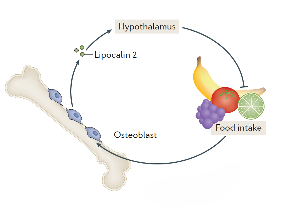
This gives us another bone–metabolism axis:
Osteoblasts → lipocalin-2 → brain appetite pathways → food intake and metabolic regulation
In diabetes and obesity, LCN2 can rise as part of inflammatory, metabolic, or renal stress. Some experimental and translational evidence suggests that increased LCN2 may act as a compensatory protective response during worsening glucose metabolism, supporting β-cell function and counteracting metabolic dysregulation26. However, chronically elevated LCN2 can also reflect tissue injury, adipose dysfunction, inflammation, or diabetic kidney disease,where NGAL/LCN2 has been studied as an early biomarker of renal tubular injury27. Therefore, LCN2 is not simply “good” or “bad”; it is a context-dependent stress and metabolic signal.
Why falls matter as much as bone strength
Fractures occur when fragile bone meets mechanical force. In diabetes, both sides of this equation are affected: the skeleton may become mechanically weaker, and the person may also become more likely to fall. The ADA Standards of Care note that fracture risk is increased in people with diabetes and is particularly relevant in those with complications such as retinopathy, neuropathy, nephropathy, poor physical function, severe hypoglycaemia, and frequent falls28.
Bone quality is reduced, but fall risk is also increased. Peripheral neuropathy can impair sensation, proprioception, balance, and gait; retinopathy can reduce visual input; hypoglycaemia can cause dizziness, confusion, or collapse; and autonomic neuropathy may contribute to orthostatic hypotension28,29. Sarcopenia, frailty, reduced handgrip strength, gait difficulty, balance impairment, foot deformity, ulcers, pain, and use of walking aids can further reduce protective responses during a fall29,30.
This means diabetes increases fracture risk not only by weakening bone but also by increasing the probability of falling. Australian osteoporosis guidance emphasises that fracture risk is shaped by clinical risk factors as well as BMD, and that increasing age, previous minimal-trauma fracture, and propensity to fall are among the strongest clinical predictors of future fracture31.
The FRAX problem: when risk calculators underestimate diabetes
RAX estimates 10-year fracture probability using clinical risk factors such as age, sex, BMI, previous fracture, parental hip fracture, smoking, glucocorticoid exposure, rheumatoid arthritis, alcohol intake, secondary osteoporosis, and femoral-neck BMD when available. The problem is that standard FRAX does not directly include type 2 diabetes, even though type 2 diabetes independently increases fracture risk and may therefore cause FRAX to underestimate true skeletal risk32,33
This is why clinicians and researchers sometimes use “the rheumatoid arthritis input as a surrogate.” It does not mean the person has rheumatoid arthritis. It means the RA checkbox is used as an imperfect proxy to increase the estimated fracture risk because diabetes adds risk that FRAX may otherwise miss. Studies and expert discussions have compared several diabetes-adjustment approaches, including using the rheumatoid arthritis input as a proxy, reducing the femoral-neck T-score by 0.5 SD, increasing the age input by 10 years, and using trabecular bone score-adjusted FRAX34,35.
The practical message is that a “low” or “borderline” FRAX result should be interpreted cautiously in people with diabetes, especially when disease duration is long or complications such as neuropathy, retinopathy, kidney disease, falls, or hypoglycaemia are present. FRAXplus now explicitly recognises type 2 diabetes as an adjustment issue and notes that diabetes duration can be incorporated into adjusted estimates, reflecting the move toward more diabetes-aware fracture prediction.
Medication choice can also influence bone risk.
Medication choice can also influence bone risk in diabetes. This does not mean glucose-lowering medicines should be avoided; rather, bone health should be considered as part of individualised diabetes care, especially in older adults, people with previous fractures, recurrent falls, neuropathy, retinopathy, kidney disease, or hypoglycaemia risk28.
Thiazolidinediones, such as pioglitazone and rosiglitazone, activate PPAR-γ (Figure 1). This can shift mesenchymal stem-cell differentiation away from osteoblast formation and toward adipocyte formation, reducing bone formation and contributing to bone loss and fracture risk36,37. In a large JAMA Internal Medicine cohort study,thiazolidinedione use was associated with increased peripheral fracture risk compared with sulfonylureas, and pioglitazone showed a stronger fracture association in that analysis37.
SGLT2 inhibitors have been questioned because of earlier concerns around canagliflozin, but more recent systematic reviews and meta-analyses generally have not shown a consistent class-wide increase in fracture risk. A network meta-analysis of 177 randomised trials found that SGLT2 inhibitors, DPP-4 inhibitors, and GLP-1 receptor agonists were not associated with increased total fracture risk in people with type 2 diabetes, while an updated SGLT2 inhibitor meta-analysis compared SGLT2 inhibitors with DPP-4 inhibitors and did not support a clear overall fracture-risk signal38,39.
GLP-1 receptor agonists are also being studied in relation to bone. Current evidence does not show a consistent increase in fracture risk in type 2 diabetes, and some meta-analytic data even suggest possible fracture-risk reduction, although conclusions remain cautious because effects may differ by drug, treatment duration, weight loss, baseline fracture risk, muscle preservation, nutrition, and physical activity40–42.
Insulin and sulfonylureas are not usually considered directly toxic to bone in the same way as thiazolidinediones, but they can increase fracture risk indirectly by increasing the risk of hypoglycaemia. In an older person, hypoglycaemia can cause dizziness, confusion, collapse, and falls. The 2026 ADA Standards of Care note that sulfonylurea use is associated with heightened fracture risk partly through hypoglycaemia-related falls, and insulin use is associated with higher hip-fracture risk, likely reflecting hypoglycaemia risk, longer diabetes duration, and comorbidities that weaken bone or increase falls1
A practical clinical framework
A patient with diabetes should be considered for skeletal risk review when they have any of the following: long duration of diabetes; type 1 diabetes beginning early in life; type 2 diabetes with neuropathy, retinopathy, chronic kidney disease, recurrent falls, or frailty; previous minimal-trauma fracture; height loss, kyphosis, or suspected vertebral fracture; use of thiazolidinediones, glucocorticoids, aromatase inhibitors, androgen-deprivation therapy, or other bone-affecting drugs; older age, low body weight, smoking, excess alcohol, poor nutrition, vitamin D deficiency, or sarcopenia1,31,35.
Assessment may include DXA, vertebral fracture assessment or spine imaging when indicated, FRAX or Garvan Fracture Risk Calculator, medication review, falls assessment, and laboratory testing such as serum calcium, phosphate, alkaline phosphatase, renal function, and 25-hydroxyvitamin D, with further investigations guided by clinical context31,43,44.
Management should be individualised, but the broad principles are consistent: optimise glycaemic control while avoiding hypoglycaemia; avoid unnecessary bone-harming drugs; support resistance, balance, and weight-bearing exercise; maintain adequate protein, calcium, and vitamin D; treat neuropathy, visual impairment, postural hypotension, and foot problems; reduce falls risk at home and in daily activity; and use osteoporosis pharmacotherapy when indicated by fracture history, BMD, or absolute fracture risk1,31,45.
The modern understanding: bone as a diabetic target organ
The most important conceptual shift is this: bone should not be seen as separate from diabetes. It is part of the metabolic network. Contemporary reviews describe diabetic bone fragility as a systemic process involving bone cells, bone matrix, vascular biology, immune-metabolic signalling, muscle, falls, and antidiabetic medication effects rather than a simple reduction in bone mineral density alone4,9.
Diabetes affects bone through hyperglycaemia, insulin deficiency or resistance, advanced glycation end products, oxidative stress, inflammation, microvascular disease, kidney disease, altered mineral metabolism, reduced bone turnover, muscle weakness, falls, and medication effects1,4.Bone also talks back to metabolism through endocrine signals such as osteocalcin and lipocalin-2, which can influence insulin secretion,insulin sensitivity, appetite regulation, energy expenditure, and broader systemic physiology14,24,26.
So the diabetic bone paradox is not merely a technical problem in DXA interpretation. It is a biological story about a skeleton that looks dense but has lost some of its material intelligence — its capacity to remodel, communicate, flex, and repair. This is why fracture risk in diabetes must be understood as a problem of bone quality, metabolic signalling, tissue repair, and fall susceptibility, not just bone quantity3,4.
Conclusion
Diabetes should no longer be viewed solely as a disorder of blood glucose regulation. It is a systemic disease that reshapes tissue integrity, vascular function, neural control, hormonal signalling, immune-metabolic balance, and the body’s capacity for repair. Within this wider biological network, the skeleton is an important but often under-recognised target.
The first fracture may appear sudden, but diabetic bone fragility usually develops silently over many years. Chronic hyperglycaemia stiffens collagen through advanced glycation, osteoblast function becomes impaired, bone remodelling slows, and microdamage accumulates. At the same time, altered osteocalcin signalling, lipocalin-2–linked metabolic stress, neuropathy, visual impairment, hypoglycaemia, sarcopenia, and falls all converge to increase fracture risk. Standard tools such as DXA and FRAX remain useful, but they may underestimate risk when bone quality, metabolic complications, and fall susceptibility are not fully considered.
The future of diabetes care should therefore include bone health as part of routine complication assessment. Alongside the eyes, kidneys, nerves, heart, and blood vessels, clinicians should also think of the skeleton. For people living with diabetes, the goal is not only better glycaemic control, but also preserved mobility, independence, fracture prevention, and long-term skeletal resilience.